
MOLE MAPPING TRACKS ANY CHANGES TO YOUR MOLES OVER TIME
London Dermatology | 3 March 2025
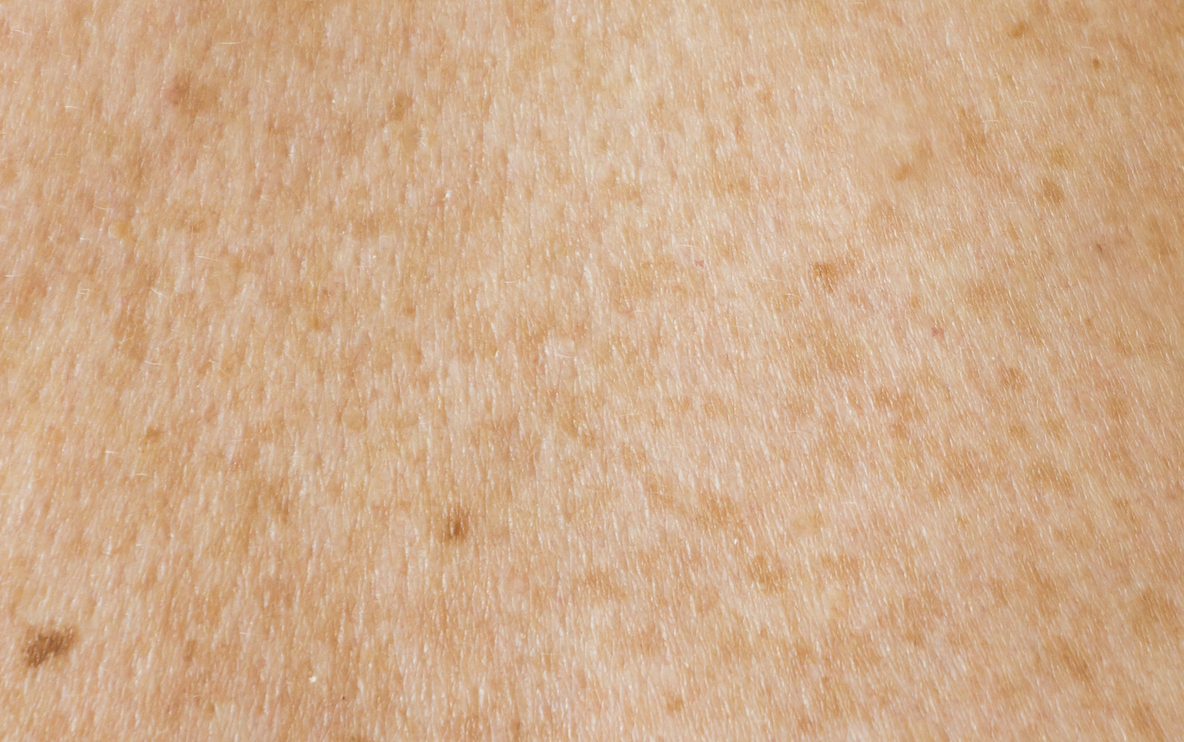
Dermatologists frequently call moles “pigmented lesions,” which are growths or patches on the skin brought on by certain skin cells known as melanocytes.
The pigment that gives the skin its colour, melanin, is made by melanocyte cells. The darker the skin or skin lesion, the more melanin there is.
Dermatologists utilise mole mapping, a trustworthy medical procedure, to track pigmented skin lesions to determine whether they are developing into cancer or have the potential to do so in the future.
Although pigmented lesions include freckles, birthmarks, and age spots, mole mapping often examines moles.
Your moles’ changes over time are tracked using mole mapping. These alterations may include modifications to the size, form, and colour of your moles as well as indications that a mole may be developing cancer.
Although mole mapping doesn’t actually prevent skin cancer, it does make it possible to diagnose the disease early, increasing the likelihood that treatment will be effective and improving patient outcomes.
The UK’s melanoma skin cancer survival rates are steadily rising, according to Cancer Research UK. If melanoma skin cancer is detected in its earliest stage, 100% of patients will survive for a year or longer after treatment, however 53% of cases are detected in its most advanced state.